Anatomy Of Chest - Female Chest Anatomy Stockfotos Und Bilder Kaufen Alamy / This thoracic and pulmonary anatomy tool is especially designed for students of anatomy (medical and paramedical studies).
Anatomy Of Chest - Female Chest Anatomy Stockfotos Und Bilder Kaufen Alamy / This thoracic and pulmonary anatomy tool is especially designed for students of anatomy (medical and paramedical studies).. These myotomes divide into the epimere and the hypomere. The superior thoracic aperture found superiorly and the inferior thoracic aperture. The chest or thorax region of the upper body has a number of important organs that reside within it that may present with chest pain if they become compromised in. This thoracic and pulmonary anatomy tool is especially designed for students of anatomy (medical and paramedical studies). The myotomes elongate and invade the mesoderm of the wall of the embryonic thoracic and abdominal cavities.
Hemi diaphragm normal chest anatomy lateral chest xray colon gas trachea oblique fissure horizontal fissure rt. See chest anatomy stock video clips. It provides protection to vital organs (eg, heart and major vessels, lungs, liver) and provides stability for movement. It also serves as a connection point for other bones and muscles. These myotomes divide into the epimere and the hypomere.
Diagram Wiring Diagram Chest Zer Full Version Hd Quality Chest Zer Activediagram Carnevalediverona It from www.amazecraze.com The chest or thorax is the region between the neck and diaphragm that encloses organs, such as the heart, lungs, esophagus, trachea, and thoracic diaphragm. Chest muscles anatomy (1) pectoralis major muscle. Sternocleidomastoid muscle clavicle and ribs anatomy muscle anatomy chest sternocleidomastoid ribs anatomy chest muscles anatomy thorax rib muscles chest muscles chest anatomy illustration. The chest is the area of origin for many of the body's systems as it houses organs such as the heart, esophagus, trachea, lungs, and thoracic diaphragm. Your sternum is a flat bone in the middle of your chest that protects the organs of your torso from injury. 30 lines of the thoracic wall syllabus p. Radiology basics of chest ct anatomy with annotated coronal images and scrollable axial images to help medical students and junior doctors learning anatomy. Thoracic cavity, also called chest cavity, the second largest hollow space of the body.
It is enclosed by the ribs, the vertebral column, and the sternum, or breastbone, and is separated from the abdominal cavity (the body's largest hollow space) by a muscular and membranous partition, the diaphragm.
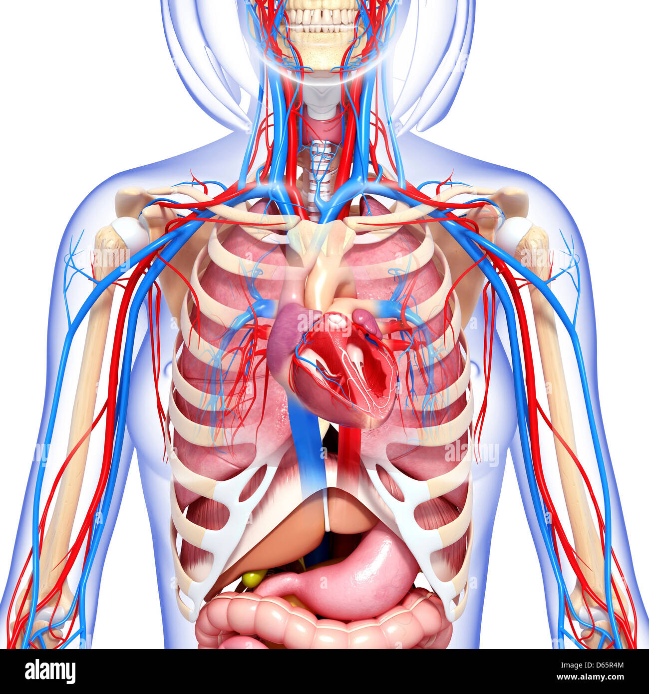
Anatomy of the chest and stomach, human anatomy, anatomy of the chest and stomach. This atlas is a comprehensive and affordable learning tool for medical students and residents and especially for radiologists and pneumologists. Your sternum is a flat bone in the middle of your chest that protects the organs of your torso from injury. The chest wall is comprised of skin, fat, muscles, and the thoracic skeleton. Plus, how to target each to make them bigger and stronger. Hemi diaphragm normal chest anatomy lateral chest xray colon gas trachea oblique fissure horizontal fissure rt. Thoracic wall the first step in understanding thorax anatomy is to find out its boundaries. Chest muscles anatomy (1) pectoralis major muscle. Anatomy of the chest and the lungs: Related posts of anatomy of the chest area anatomy of the female human body. Learn about each of these muscles, their locations, functional anatomy and exercises for them. See human chest anatomy stock video clips. 31 anatomy of the female breast syllabus p.
Anatomy of the chest and the lungs: The epidermis is the outermost layer that provides a protective, waterproof seal over the body. Most people struggle to build the top portion of their chest, so we'll pay special attention to this area. This thoracic and pulmonary anatomy tool is especially designed for students of anatomy (medical and paramedical studies). The pectoralis major and the pectoralis minor, known collectively as your pecs.
Female Chest Anatomy Stockfotos Und Bilder Kaufen Alamy from c8.alamy.com Large, complex chest wall defects can be some of the most challenging problems a reconstructive surgeon must face, but successful outcomes may be reliably achieved by adhering to basic principles of adequate debridement followed by. In insects, crustaceans, and the extinct trilobites, the thorax is one of the three main divisions of the creature's body, each of which is in turn composed of multiple segments. Learn about each of these muscles, their locations, functional anatomy and exercises for them. The circulatory system does most of its work. This atlas is a comprehensive and affordable learning tool for medical students and residents and especially for radiologists and pneumologists. Computed tomography (ct) of the chest can detect pathology that may not show up on a conventional chest radiograph(1). #anatomy of the chest and stomach. It provides protection to vital organs (eg, heart and major vessels, lungs, liver) and provides stability for movement.
This thoracic and pulmonary anatomy tool is especially designed for students of anatomy (medical and paramedical studies).
System respiratory respiratory organs of human body digestive and respiratory system medical chest internal structure of human body medicine body lungs biology intestines stomach anatomy torso human internal. Plus, how to target each to make them bigger and stronger. The muscles of the chest develop from the somites found in the mesoderm. A line is drawn from anterior surface of the body of 6th thoracic vertebrae passing through the apex of the heart up to anterior lower most part of diaphragm. In insects, crustaceans, and the extinct trilobites, the thorax is one of the three main divisions of the creature's body, each of which is in turn composed of multiple segments. The chest wall is comprised of skin, fat, muscles, and the thoracic skeleton. This thoracic and pulmonary anatomy tool is especially designed for students of anatomy (medical and paramedical studies). It spreads out like a fan and covers the rib cage like an armor plate. This page provides an overview of the chest muscle group. The thorax has two major openings: Here, we break down the anatomy of your chest muscles. 30 lines of the thoracic wall syllabus p. How to view the anatomical labels.
Chest a man's chest — like the rest of his body — is covered with skin that has two layers. System respiratory respiratory organs of human body digestive and respiratory system medical chest internal structure of human body medicine body lungs biology intestines stomach anatomy torso human internal. The chest anatomy includes the pectoralis major, pectoralis minor and the serratus anterior. Computed tomography (ct) of the chest can detect pathology that may not show up on a conventional chest radiograph(1). Related posts of anatomy of the chest area anatomy of the female human body.
Model Of The Intern Anatomy Of The Chest Of An Asexual Adult Human Body Face On Stock Photo Picture And Rights Managed Image Pic Bsi 1137905 Agefotostock from previews.agefotostock.com It spreads out like a fan and covers the rib cage like an armor plate. Related posts of anatomy of the chest area anatomy of the female human body. Browse 2,911 anatomy of the chest organs stock photos and images available, or start a new search to explore more stock photos and images. It also serves as a connection point for other bones and muscles. Thoracic cavity, also called chest cavity, the second largest hollow space of the body. The upper part of your pec major, the clavicular head runs from your clavicle (collarbone) across the top of your chest and attaches to your humerus, or upper arm. The thorax has two major openings: Hemi diaphragm normal chest anatomy lateral chest xray colon gas trachea oblique fissure horizontal fissure rt.
The superior thoracic aperture found superiorly and the inferior thoracic aperture.
Thoracic cavity, also called chest cavity, the second largest hollow space of the body. These myotomes divide into the epimere and the hypomere. The circulatory system does most of its work. The ribs and sternum make up what is called the 'ribcage.' the ribcage protects the lungs, blood vessels, and heart. Related posts of anatomy of the chest area anatomy of the female human body. See human chest anatomy stock video clips. 12 photos of the anatomy of the chest and stomach. The right side of the heart is deflected anteriorly, and the left side is deflected posteriorly. 4 innervation of the breast blood supply of the breast syllabus p. Anatomy of the chest and stomach, human anatomy, anatomy of the chest and stomach. Large, complex chest wall defects can be some of the most challenging problems a reconstructive surgeon must face, but successful outcomes may be reliably achieved by adhering to basic principles of adequate debridement followed by. It is enclosed by the ribs, the vertebral column, and the sternum, or breastbone, and is separated from the abdominal cavity (the body's largest hollow space) by a muscular and membranous partition, the diaphragm. Radiology basics of chest ct anatomy with annotated coronal images and scrollable axial images to help medical students and junior doctors learning anatomy.


0 Komentar